 会员登录
会员登录 购物车()
购物车()

 成功收藏产品
商家描述
包装清单
售后服务
产品评价(0)
概述
产品描述Parkin is a zinc-finger protein that is related to ubiquitin at the amino terminus. The wild type Parkin gene, which maps to human chromosome 6q25.2-27, encodes a 465 amino acid full-length protein that is expressed as multiple isoforms. Mutations in the Parkin gene are responsible for autosomal recessive juvenile Parkinson's disease and commonly involve deletions of exons 3-5. In humans, Parkin is expressed in a subset of cells of the basal ganglia, midbrain, cerebellum and cerebral cortex, and is subject to alternative splicing in different tissues. Parkin expression is also high in the brainstem of mice, with the majority of immunopositive cells being neurons. The Parkin gene has been identified in a diverse group of organisms including mammals, birds, frog and fruit flies, suggesting that analogous functional roles of the Parkin protein may have been highly conserved during the course of evolution.
产品名称Anti-Parkin Recombinant Rabbit Monoclonal Antibody [JF82-09]
分子量52 kDa
种属反应性Human,Mouse,Rat
验证应用WB,ICC/IF,IHC-P,IP,FC
抗体类型重组兔单抗
免疫原Synthetic peptide within N-terminal human Parkin.
偶联Non-conjugated
性能
形态Liquid
浓度1 mg/mL.
存放说明Store at +4℃ after thawing. Aliquot store at -20℃ or -80℃. Avoid repeated freeze / thaw cycles.
存储缓冲液1*TBS (pH7.4), 0.05% BSA, 40% Glycerol. Preservative: 0.05% Sodium Azide.
亚型IgG
纯化方式Protein A affinity purified.
亚细胞定位Mitochondrion, mitochondrion outer membrane, endoplasmic reticulum, cytosol, Nucleus, neuron projection, postsynaptic density, presynapse.
数据链接SwissProt: O60260 Human
SwissProt: Q9WVS6 Mouse
SwissProt: Q9JK66 Rat
其它名称
AR JP antibody
E3 ubiquitin ligase antibody
E3 ubiquitin protein ligase parkin antibody
more
应用
WB: 1:500-1:1,000
ICC/IF: 1:50-1:200
IHC-P: 1:50-1:200
FC: 1:50-1:100
IP: Use at an assay dependent concentration.
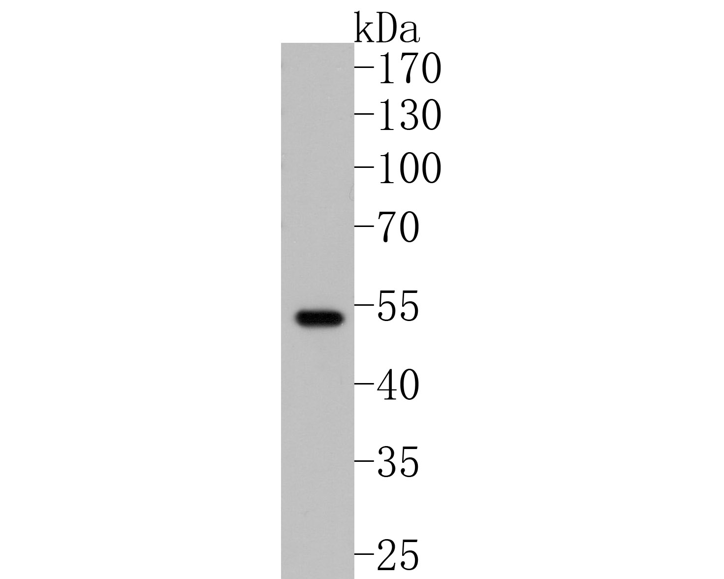
Fig1: Western blot analysis of Parkin on 293T cell lysates. Proteins were transferred to a PVDF membrane and blocked with 5% BSA in PBS for 1 hour at room temperature. The primary antibody (ET1702-60, 1/500) was used in 5% BSA at room temperature for 2 hours. Goat Anti-Rabbit IgG - HRP Secondary Antibody (HA1001) at 1:5,000 dilution was used for 1 hour at room temperature.
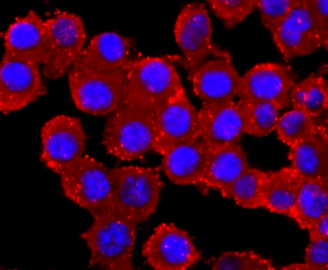
Fig2: ICC staining of Parkin in N2A cells (red). Formalin fixed cells were permeabilized with 0.1% Triton X-100 in TBS for 10 minutes at room temperature and blocked with 1% Blocker BSA for 15 minutes at room temperature. Cells were probed with the primary antibody (ET1702-60, 1/50) for 1 hour at room temperature, washed with PBS. Alexa Fluor®594 Goat anti-Rabbit IgG was used as the secondary antibody at 1/1,000 dilution. The nuclear counter stain is DAPI (blue).
Fig3: ICC staining of Parkin in SH-SY5Y cells (red). Formalin fixed cells were permeabilized with 0.1% Triton X-100 in TBS for 10 minutes at room temperature and blocked with 1% Blocker BSA for 15 minutes at room temperature. Cells were probed with the primary antibody (ET1702-60, 1/50) for 1 hour at room temperature, washed with PBS. Alexa Fluor®594 Goat anti-Rabbit IgG was used as the secondary antibody at 1/1,000 dilution. The nuclear counter stain is DAPI (blue).
Fig4: ICC staining of Parkin in PC-3M cells (red). Formalin fixed cells were permeabilized with 0.1% Triton X-100 in TBS for 10 minutes at room temperature and blocked with 1% Blocker BSA for 15 minutes at room temperature. Cells were probed with the primary antibody (ET1702-60, 1/50) for 1 hour at room temperature, washed with PBS. Alexa Fluor®594 Goat anti-Rabbit IgG was used as the secondary antibody at 1/1,000 dilution. The nuclear counter stain is DAPI (blue).
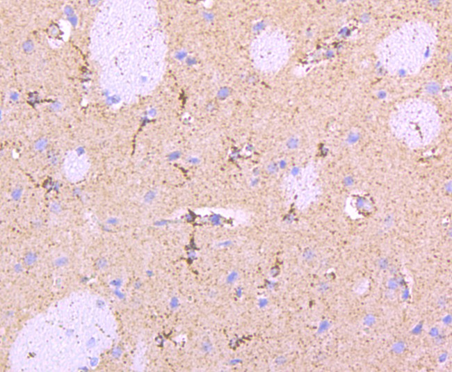
Fig5: Immunohistochemical analysis of paraffin-embedded rat brain tissue using anti-Parkin antibody. The section was pre-treated using heat mediated antigen retrieval with Tris-EDTA buffer (pH 8.0-8.4) for 20 minutes.The tissues were blocked in 5% BSA for 30 minutes at room temperature, washed with ddH2O and PBS, and then probed with the primary antibody (ET1702-60, 1/50) for 30 minutes at room temperature. The detection was performed using an HRP conjugated compact polymer system. DAB was used as the chromogen. Tissues were counterstained with hematoxylin and mounted with DPX.
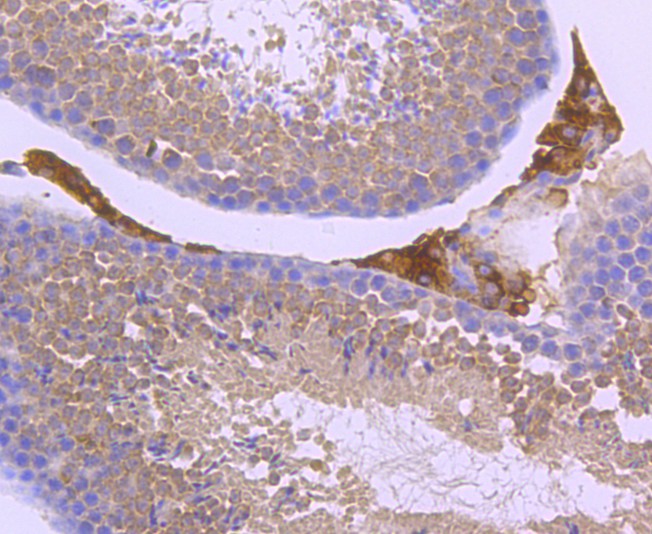
Fig6: Immunohistochemical analysis of paraffin-embedded mouse testis tissue using anti-Parkin antibody. The section was pre-treated using heat mediated antigen retrieval with Tris-EDTA buffer (pH 8.0-8.4) for 20 minutes.The tissues were blocked in 5% BSA for 30 minutes at room temperature, washed with ddH2O and PBS, and then probed with the primary antibody (ET1702-60, 1/50) for 30 minutes at room temperature. The detection was performed using an HRP conjugated compact polymer system. DAB was used as the chromogen. Tissues were counterstained with hematoxylin and mounted with DPX.
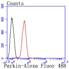
Fig7: Flow cytometric analysis of Parkin was done on SH-SY5Y cells. The cells were fixed, permeabilized and stained with the primary antibody (ET1702-60, 1/50) (red). After incubation of the primary antibody at room temperature for an hour, the cells were stained with a Alexa Fluor 488-conjugated Goat anti-Rabbit IgG Secondary antibody at 1/1000 dilution for 30 minutes.Unlabelled sample was used as a control (cells without incubation with primary antibody; black).
背景文献
1. Lu Y et al. Beneficial effects of astragaloside IV against angiotensin II-induced mitochondrial dysfunction in rat vascular smooth muscle cells. Int J Mol Med 36:1223-32 (2015).
2. Seillier M et al. Defects in mitophagy promote redox-driven metabolic syndrome in the absence of TP53INP1. EMBO Mol Med 7:802-18 (2015).