产品描述Autophagy receptor required for selective macroautophagy (aggrephagy). Functions as a bridge between polyubiquitinated cargo and autophagosomes. Interacts directly with both the cargo to become degraded and an autophagy modifier of the MAP1 LC3 family. Along with WDFY3, involved in the formation and autophagic degradation of cytoplasmic ubiquitin-containing inclusions (p62 bodies, ALIS/aggresome-like induced structures). Along with WDFY3, required to recruit ubiquitinated proteins to PML bodies in the nucleus. May regulate the activation of NFKB1 by TNF-alpha, nerve growth factor (NGF) and interleukin-1. May play a role in titin/TTN downstream signaling in muscle cells. May regulate signaling cascades through ubiquitination. Adapter that mediates the interaction between TRAF6 and CYLD. May be involved in cell differentiation, apoptosis, immune response and regulation of K+ channels. Involved in endosome organization by retaining vesicles in the perinuclear cloud: following ubiquitination by RNF26, attracts specific vesicle-associated adapters, forming a molecular bridge that restrains cognate vesicles in the perinuclear region and organizes the endosomal pathway for efficient cargo transport. Promotes relocalization of 'Lys-63'-linked ubiquitinated STING1 to autophagosomes. Acts as an activator of the NFE2L2/NRF2 pathway via interaction with KEAP1: interaction inactivates the BCR(KEAP1) complex, promoting nuclear accumulation of NFE2L2/NRF2 and subsequent expression of cytoprotective genes.
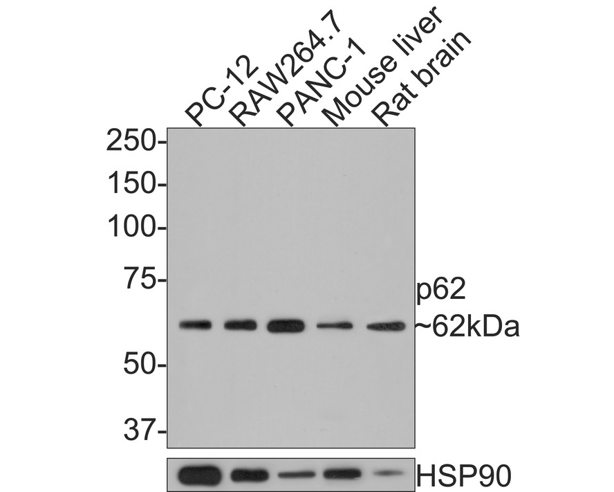
Fig1: Western blot analysis of p62 on different lysates with Rabbit anti-p62 antibody (HA721171) at 1/500 dilution.
Lane 1: PC-12 cell lysate (10 µg/Lane)
Lane 2: RAW264.7 cell lysate (10 µg/Lane)
Lane 3: PANC-1 cell lysate (10 µg/Lane)
Lane 4: Mouse liver tissue lysate (20 µg/Lane)
Lane 5: Rat brain tissue lysate (20 µg/Lane)
Predicted band size: 48 kDa
Observed band size: 62 kDa
Exposure time: 2 minutes;
8% SDS-PAGE gel.
Proteins were transferred to a PVDF membrane and blocked with 5% NFDM/TBST for 1 hour at room temperature. The primary antibody (HA721171) at 1/500 dilution was used in 5% NFDM/TBST at room temperature for 2 hours. Goat Anti-Rabbit IgG - HRP Secondary Antibody (HA1001) at 1:300,000 dilution was used for 1 hour at room temperature.
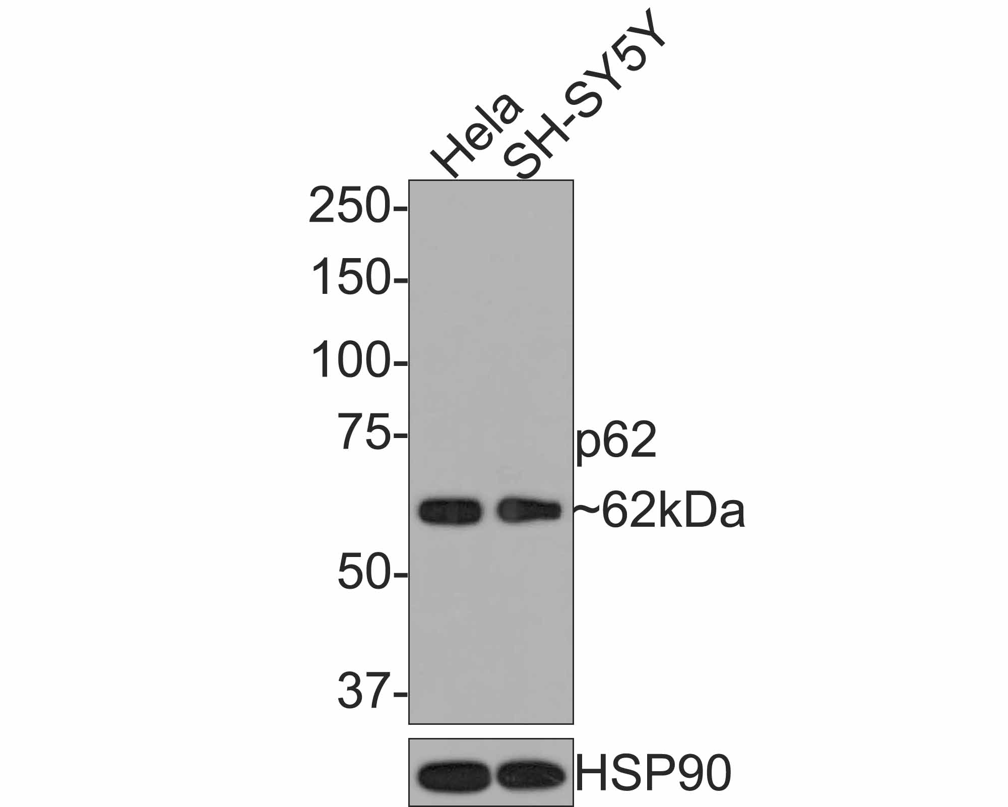
Fig2: Western blot analysis of p62 on different lysates with Rabbit anti-p62 antibody (HA721171) at 1/1,000 dilution.
Lane 1: Hela cell lysate
Lane 2: SH-SY5Y cell lysate
Lysates/proteins at 10 µg/Lane.
Predicted band size: 48 kDa
Observed band size: 62 kDa
Exposure time: 2 minutes;
8% SDS-PAGE gel.
Proteins were transferred to a PVDF membrane and blocked with 5% NFDM/TBST for 1 hour at room temperature. The primary antibody (HA721171) at 1/1,000 dilution was used in 5% NFDM/TBST at room temperature for 2 hours. Goat Anti-Rabbit IgG - HRP Secondary Antibody (HA1001) at 1:300,000 dilution was used for 1 hour at room temperature.
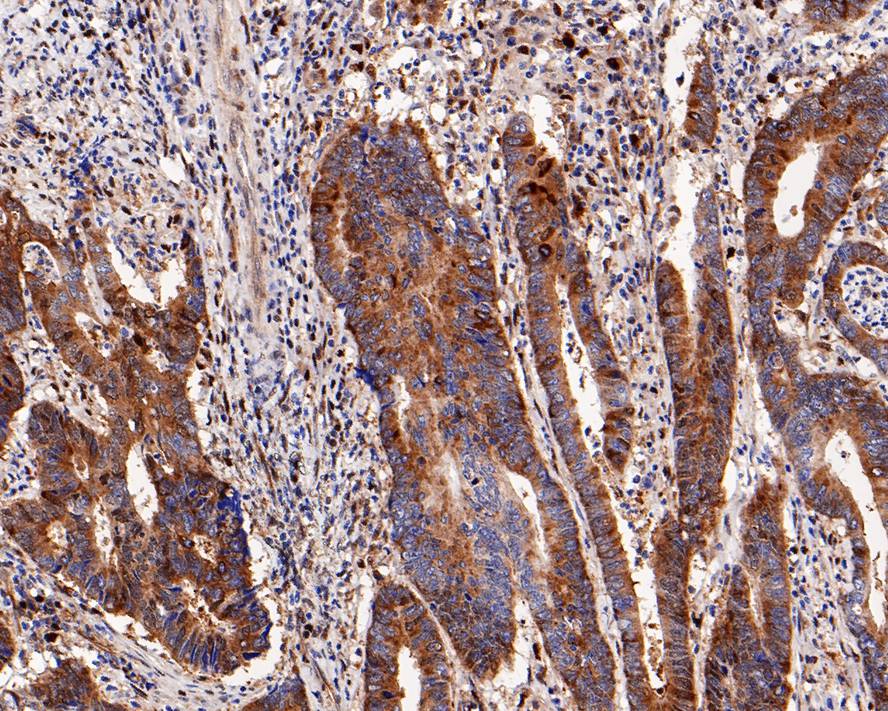
Fig3: Immunohistochemical analysis of paraffin-embedded human colon carcinoma tissue with Rabbit anti-p62 antibody (HA721171) at 1/400 dilution.
The section was pre-treated using heat mediated antigen retrieval with sodium citrate buffer (pH 6.0) for 2 minutes. The tissues were blocked in 1% BSA for 20 minutes at room temperature, washed with ddH2O and PBS, and then probed with the primary antibody (HA721171) at 1/400 dilution for 1 hour at room temperature. The detection was performed using an HRP conjugated compact polymer system. DAB was used as the chromogen. Tissues were counterstained with hematoxylin and mounted with DPX.
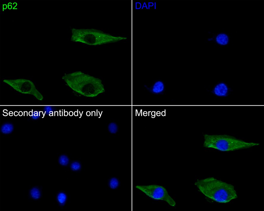
Fig4: Immunocytochemistry analysis of A549 cells labeling p62 with Rabbit anti-p62 antibody (HA721171) at 1/50 dilution.
Cells were fixed in 4% paraformaldehyde for 10 minutes at 37 ℃, permeabilized with 0.05% Triton X-100 in PBS for 20 minutes, and then blocked with 2% negative goat serum for 30 minutes at room temperature. Cells were then incubated with Rabbit anti-p62 antibody (HA721171) at 1/50 dilution in 2% negative goat serum overnight at 4 ℃. Goat Anti-Rabbit IgG H&L (iFluor™ 488, HA1121) was used as the secondary antibody at 1/1,000 dilution. PBS instead of the primary antibody was used as the secondary antibody only control. Nuclear DNA was labelled in blue with DAPI.
背景文献
1. Tan C.T., Chang H.C., Zhou Q., Yu C., Fu N.Y., Sabapathy K., Yu V.C. MOAP-1-mediated dissociation of p62/SQSTM1 bodies releases Keap1 and suppresses Nrf2 signaling. EMBO Rep. 22:e50854-e50854(2021)
2. Prabakaran T., Bodda C., Krapp C., Zhang B.C., Christensen M.H., Sun C., Reinert L., Cai Y., Jensen S.B., Paludan S.R. Attenuation of cGAS-STING signaling is mediated by a p62/SQSTM1-dependent autophagy pathway activated by TBK1. EMBO J. 37:0-0(2018)
 会员登录
会员登录 购物车()
购物车()

 成功收藏产品


