产品描述Gasdermin-D, N-terminal: Promotes pyroptosis in response to microbial infection and danger signals. Produced by the cleavage of gasdermin-D by inflammatory caspases CASP1 or CASP4 in response to canonical, as well as non-canonical (such as cytosolic LPS) inflammasome activators. After cleavage, moves to the plasma membrane where it strongly binds to inner leaflet lipids, including monophosphorylated phosphatidylinositols, such as phosphatidylinositol 4-phosphate, bisphosphorylated phosphatidylinositols, such as phosphatidylinositol (4,5)-bisphosphate, as well as phosphatidylinositol (3,4,5)-bisphosphate, and more weakly to phosphatidic acid and phosphatidylserine. Homooligomerizes within the membrane and forms pores of 10 - 15 nanometers (nm) of inner diameter, possibly allowing the release of mature IL1B and triggering pyroptosis . Exhibits bactericidal activity. Gasdermin-D, N-terminal released from pyroptotic cells into the extracellular milieu rapidly binds to and kills both Gram-negative and Gram-positive bacteria, without harming neighboring mammalian cells, as it does not disrupt the plasma membrane from the outside due to lipid-binding specificity.
产品名称Anti-Gasdermin D (N terminal) Recombinant Rabbit Monoclonal Antibody [PD00-18]
分子量Predicted band size: 53/30 kDa
种属反应性Human,Mouse,Rat
验证应用WB
抗体类型重组兔单抗
免疫原Recombinant protein within Gasdermin D full length protein.
偶联Non-conjugated
性能
形态Liquid
浓度1 mg/mL.
存放说明Store at +4℃ after thawing. Aliquot store at -20℃. Avoid repeated freeze / thaw cycles.
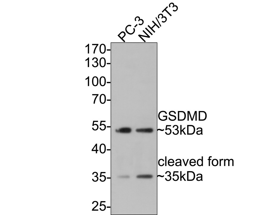
Fig1: Western blot analysis of Gasdermin D (N terminal) on different lysates with Rabbit anti-Gasdermin D (N terminal) antibody (HA721144) at 1/500 dilution.
Lane 1: PC-3 cell lysate
Lane 2: NIH/3T3 cell lysate
Lysates/proteins at 10 µg/Lane.
Predicted band size: 53 kDa
Observed band size: 53/35 kDa
Exposure time: 30 seconds;
10% SDS-PAGE gel.
Proteins were transferred to a PVDF membrane and blocked with 5% NFDM/TBST for 1 hour at room temperature. The primary antibody (HA721144) at 1/500 dilution was used in 5% NFDM/TBST at room temperature for 2 hours. Goat Anti-Rabbit IgG - HRP Secondary Antibody (HA1001) at 1:300,000 dilution was used for 1 hour at room temperature.
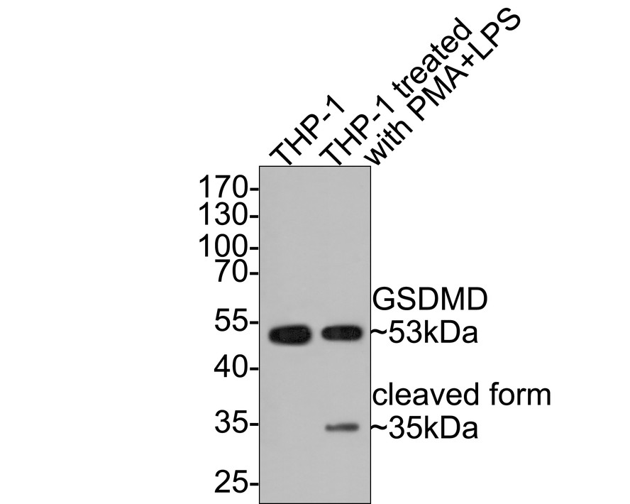
Fig2: Western blot analysis of Gasdermin D (N terminal) on different lysates with Rabbit anti-Gasdermin D (N terminal) antibody (HA721144) at 1/500 dilution.
Lane 1: THP-1 cell lysate
Lane 2: THP-1 cell lysate treated with PMA for 17.5 hours and then treated with LPS for 6 hours
Lysates/proteins at 10 µg/Lane.
Predicted band size: 53 kDa
Observed band size: 53/35 kDa
Exposure time: 30 seconds;
10% SDS-PAGE gel.
Proteins were transferred to a PVDF membrane and blocked with 5% NFDM/TBST for 1 hour at room temperature. The primary antibody (HA721144) at 1/500 dilution was used in 5% NFDM/TBST at room temperature for 2 hours. Goat Anti-Rabbit IgG - HRP Secondary Antibody (HA1001) at 1:300,000 dilution was used for 1 hour at room temperature.
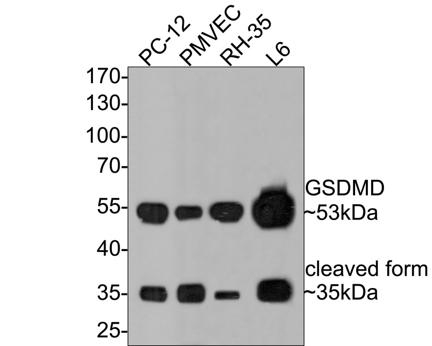
Fig3: Western blot analysis of Gasdermin D (N terminal) on different lysates with Rabbit anti-Gasdermin D (N terminal) antibody (HA721144) at 1/500 dilution.
Lane 1: PC-12 cell lysate
Lane 2: PMVEC cell lysate
Lane 3: RH-35 cell lysate
Lane 4: L6 cell lysate
Lysates/proteins at 10 µg/Lane.
Predicted band size: 53 kDa
Observed band size: 53/35 kDa
Exposure time: 2 minutes;
10% SDS-PAGE gel.
Proteins were transferred to a PVDF membrane and blocked with 5% NFDM/TBST for 1 hour at room temperature. The primary antibody (HA721144) at 1/500 dilution was used in 5% NFDM/TBST at room temperature for 2 hours. Goat Anti-Rabbit IgG - HRP Secondary Antibody (HA1001) at 1:300,000 dilution was used for 1 hour at room temperature.
背景文献
1. Sborgi L. et. al. GSDMD membrane pore formation constitutes the mechanism of pyroptotic cell death. EMBO J. 35:1766-1778(2016).
2. Ding J. et. al. Pore-forming activity and structural autoinhibition of the gasdermin family. Nature 535:111-116(2016).
 会员登录
会员登录 购物车()
购物车()

 成功收藏产品


