 会员登录
会员登录 购物车()
购物车()

- 商城价格 登录后可查看价格
- 货号 ET1610-93
- 品牌 huabio/华安生物 ( 经销商 )
- CAS号
- 规格/包装 50uL
- 单位 支
- 储存条件 -20℃
- 现货状态 一周
-
数量
- +
 成功收藏产品
 - 立即购买
 热门推荐
热门推荐
-
概述
- 产品描述NLRP3 is expressed predominantly in macrophages and as a component of the inflammasome, detects products of damaged cells such as extracellular ATP and crystalline uric acid. Activated NLRP3 in turn triggers an immune response. Mutations in the NLRP3 gene are associated with a number of organ specific autoimmune diseases. NLRP3 is a component of the innate immune system that functions as a pattern recognition receptor (PRR) that recognizes pathogen-associated molecular patterns (PAMPs). NLRP3 belongs to the NOD-like receptor (NLR) subfamily of PRRs and NLRP3 together with the adaptor ASC protein PYCARD forms a caspase-1 activating complex known as the NLRP3 inflammasome. NLRP3 in the absence of activating signal is kept in an inactive state complexed with HSP90 and SGT1 in the cytoplasm. NLRP3 inflammasome detects danger signals such as crystalline uric acid and extracellular ATP released by damaged cells. These signals release HSP90 and SGT1 from and recruit ASC protein and caspase-1 to the inflammasome complex. Caspase-1 within the activated NLRP3 inflammasome complex in turn activates the inflammatory cytokine, IL-1β. The NLRP3 inflammasome appears to be activated by changes in intracellular potassium caused by potassium efflux from mechanosensitive ion channels located in the cell membrane
- 产品名称Anti-NLRP3 Recombinant Rabbit Monoclonal Antibody [SC06-23]
- 分子量Predicted band size: 118 kDa
- 种属反应性Human,Mouse,Rat
- 验证应用WB,ICC/IF,IHC-P,FC,IP
- 抗体类型重组兔单抗
- 免疫原Recombinant protein within Human NLRP3 aa 5-161 / 1036.
- 偶联Non-conjugated
-
性能
- 形态Liquid
- 浓度1 mg/mL.
- 存放说明Store at +4℃ after thawing. Aliquot store at -20℃ or -80℃. Avoid repeated freeze / thaw cycles.
- 存储缓冲液1*TBS (pH7.4), 0.05% BSA, 40% Glycerol. Preservative: 0.05% Sodium Azide.
- 亚型IgG
- 纯化方式Protein A affinity purified.
- 亚细胞定位Cytoplasm, Inflammasome, Secreted, Nucleus, Endoplasmic reticulum.
-
数据链接SwissProt: Q96P20 Human
SwissProt: Q8R4B8 Mouse
Entrez Gene: 287362 Rat
-
其它名称
- AGTAVPRL antibody
- AII/AVP antibody
- Angiotensin/vasopressin receptor AII/AVP like antibody
-
应用
WB: 1:500-1:1,000
ICC/IF: 1:100-1:500
IHC-P: 1:50-1:400
FC: 1:50-1:100
IP: Use at an assay dependent concentration.
-

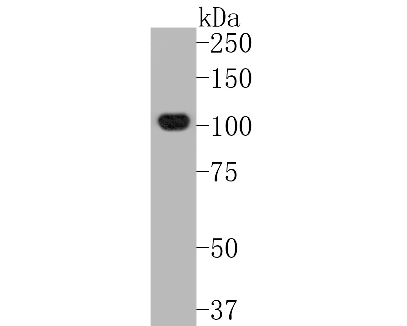
Fig1: Western blot analysis of NLRP3 on human lung tissue lysates. Proteins were transferred to a PVDF membrane and blocked with 5% BSA in PBS for 1 hour at room temperature. The primary antibody (ET1610-93, 1/500) was used in 5% BSA at room temperature for 2 hours. Goat Anti-Rabbit IgG - HRP Secondary Antibody (HA1001) at 1:5,000 dilution was used for 1 hour at room temperature.

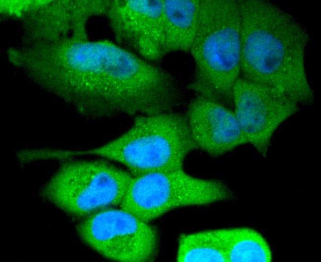
Fig2: ICC staining of NLRP3 in Hela cells (green). Formalin fixed cells were permeabilized with 0.1% Triton X-100 in TBS for 10 minutes at room temperature and blocked with 1% Blocker BSA for 15 minutes at room temperature. Cells were probed with the primary antibody (ET1610-93, 1/50) for 1 hour at room temperature, washed with PBS. Alexa Fluor®488 Goat anti-Rabbit IgG was used as the secondary antibody at 1/1,000 dilution. The nuclear counter stain is DAPI (blue).

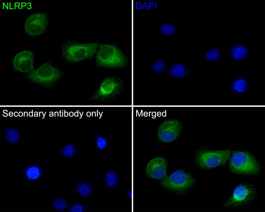
Fig3: Immunocytochemistry analysis of HUVEC cells labeling NLRP3 with Rabbit anti-NLRP3 antibody (ET1610-93) at 1/50 dilution.
Cells were fixed in 4% paraformaldehyde for 10 minutes at 37 ℃, permeabilized with 0.05% Triton X-100 in PBS for 20 minutes, and then blocked with 2% negative goat serum for 30 minutes at room temperature. Cells were then incubated with Rabbit anti-NLRP3 antibody (ET1610-93) at 1/50 dilution in 2% negative goat serum overnight at 4 ℃. Goat Anti-Rabbit IgG H&L (iFluor™ 488, HA1121) was used as the secondary antibody at 1/1,000 dilution. PBS instead of the primary antibody was used as the secondary antibody only control. Nuclear DNA was labelled in blue with DAPI.
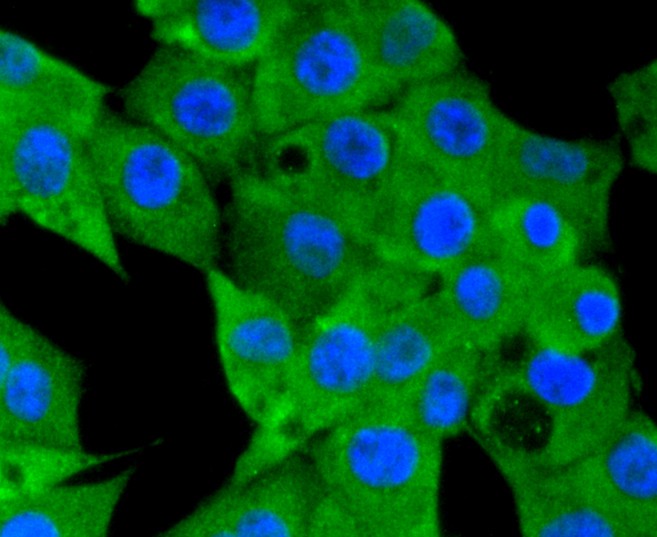
Fig4: ICC staining of NLRP3 in PMVEC cells (green). Formalin fixed cells were permeabilized with 0.1% Triton X-100 in TBS for 10 minutes at room temperature and blocked with 1% Blocker BSA for 15 minutes at room temperature. Cells were probed with the primary antibody (ET1610-93, 1/50) for 1 hour at room temperature, washed with PBS. Alexa Fluor®488 Goat anti-Rabbit IgG was used as the secondary antibody at 1/1,000 dilution. The nuclear counter stain is DAPI (blue).

Fig5: Immunohistochemical analysis of paraffin-embedded mouse bladder tissue using anti-NLRP3 antibody. The section was pre-treated using heat mediated antigen retrieval with Tris-EDTA buffer (pH 9.0) for 20 minutes.The tissues were blocked in 1% BSA for 30 minutes at room temperature, washed with ddH2O and PBS, and then probed with the primary antibody (ET1610-93, 1/50) for 30 minutes at room temperature. The detection was performed using an HRP conjugated compact polymer system. DAB was used as the chromogen. Tissues were counterstained with hematoxylin and mounted with DPX.

Fig6: Immunohistochemical analysis of paraffin-embedded mouse spleen tissue using anti-NLRP3 antibody. The section was pre-treated using heat mediated antigen retrieval with Tris-EDTA buffer (pH 9.0) for 20 minutes.The tissues were blocked in 1% BSA for 30 minutes at room temperature, washed with ddH2O and PBS, and then probed with the primary antibody (ET1610-93, 1/50) for 30 minutes at room temperature. The detection was performed using an HRP conjugated compact polymer system. DAB was used as the chromogen. Tissues were counterstained with hematoxylin and mounted with DPX.

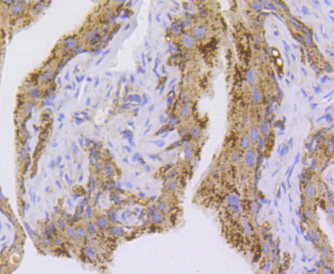
Fig7: Immunohistochemical analysis of paraffin-embedded human lung carcinoma tissue using anti-NLRP3 antibody. The section was pre-treated using heat mediated antigen retrieval with Tris-EDTA buffer (pH 9.0) for 20 minutes.The tissues were blocked in 1% BSA for 30 minutes at room temperature, washed with ddH2O and PBS, and then probed with the primary antibody (ET1610-93, 1/50) for 30 minutes at room temperature. The detection was performed using an HRP conjugated compact polymer system. DAB was used as the chromogen. Tissues were counterstained with hematoxylin and mounted with DPX.

Fig8: Flow cytometric analysis of NLRP3 was done on Jurkat cells. The cells were fixed, permeabilized and stained with the primary antibody (ET1610-93, 1/50) (red). After incubation of the primary antibody at room temperature for an hour, the cells were stained with a Alexa Fluor 488-conjugated Goat anti-Rabbit IgG Secondary antibody at 1/1,000 dilution for 30 minutes.Unlabelled sample was used as a control (cells without incubation with primary antibody; black).

Fig9: Immunohistochemical analysis of paraffin-embedded human colon carcinoma tissue with Rabbit anti-NLRP3 antibody (ET1610-93) at 1/400 dilution.
The section was pre-treated using heat mediated antigen retrieval with sodium citrate buffer (pH 6.0) for 2 minutes. The tissues were blocked in 1% BSA for 20 minutes at room temperature, washed with ddH2O and PBS, and then probed with the primary antibody (ET1610-93) at 1/400 dilution for 1 hour at room temperature. The detection was performed using an HRP conjugated compact polymer system. DAB was used as the chromogen. Tissues were counterstained with hematoxylin and mounted with DPX.
Fig10: Immunohistochemical analysis of paraffin-embedded human liver carcinoma tissue with Rabbit anti-NLRP3 antibody (ET1610-93) at 1/100 dilution.
The section was pre-treated using heat mediated antigen retrieval with sodium citrate buffer (pH 6.0) for 2 minutes. The tissues were blocked in 1% BSA for 20 minutes at room temperature, washed with ddH2O and PBS, and then probed with the primary antibody (ET1610-93) at 1/100 dilution for 1 hour at room temperature. The detection was performed using an HRP conjugated compact polymer system. DAB was used as the chromogen. Tissues were counterstained with hematoxylin and mounted with DPX.


-
背景文献
-
1. Qiao J et al. Busulfan and cyclosphamide induce liver inflammation through NLRP3 activation in mice after hematopoietic stem cell transplantation. Sci Rep 5:17828 (2015).
2. Ataide MA et al. Malaria-induced NLRP12/NLRP3-dependent caspase-1 activation mediates inflammation and hypersensitivity to bacterial superinfection. PLoS Pathog 10:e1003885 (2014).