产品描述Transmembrane glycoprotein of the endoplasmic reticulum that functions as a transcription activator and initiates the unfolded protein response (UPR) during endoplasmic reticulum stress. Cleaved upon ER stress, the N-terminal processed cyclic AMP-dependent transcription factor ATF-6 alpha translocates to the nucleus where it activates transcription of genes involved in the UPR. Binds DNA on the 5'-CCAC[GA]-3'half of the ER stress response element (ERSE) (5'-CCAAT-N9-CCAC[GA]-3') and of ERSE II (5'-ATTGG-N-CCACG-3'). Binding to ERSE requires binding of NF-Y to ERSE. Could also be involved in activation of transcription by the serum response factor. May play a role in foveal development and cone function in the retina.
产品名称Anti-ATF6 Mouse Monoclonal Antibody [8D3]
分子量Predicted band size: 75 kDa
种属反应性Human,Mouse,Rat
验证应用WB,IHC-P,FC
抗体类型小鼠单抗
免疫原Recombinant protein within Human ATF6 aa 1-670 / -670.
偶联Non-conjugated
性能
形态Liquid
浓度2 mg/mL.
存放说明Store at +4℃ after thawing. Aliquot store at -20℃. Avoid repeated freeze / thaw cycles.
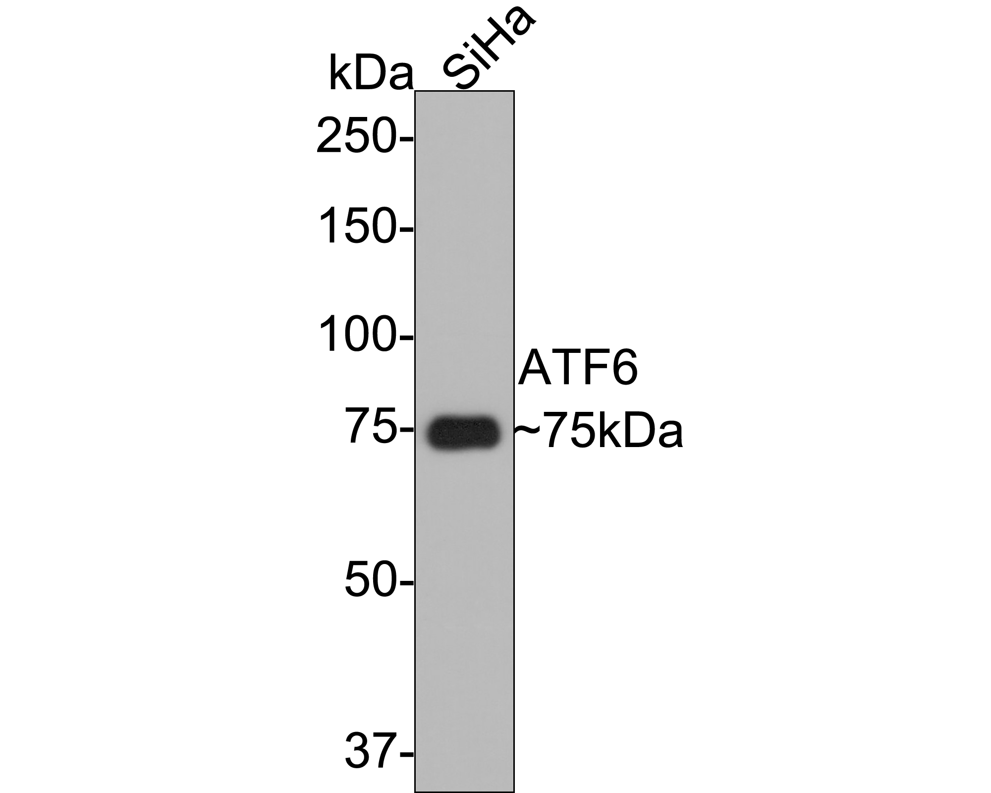
Fig1: Western blot analysis of ATF6 on SiHa cell lysates with Mouse anti-ATF6 antibody (EM1701-94) at 1/5,000 dilution.
Lysates/proteins at 10 µg/Lane.
Predicted band size: 75 kDa
Observed band size: 75 kDa
Exposure time: 30 seconds;
8% SDS-PAGE gel.
Proteins were transferred to a PVDF membrane and blocked with 5% NFDM/TBST for 1 hour at room temperature. The primary antibody (EM1701-94) at 1/5,000 dilution was used in 5% NFDM/TBST at room temperature for 2 hours. Goat Anti-Mouse IgG - HRP Secondary Antibody (HA1006) at 1:100,000 dilution was used for 1 hour at room temperature.
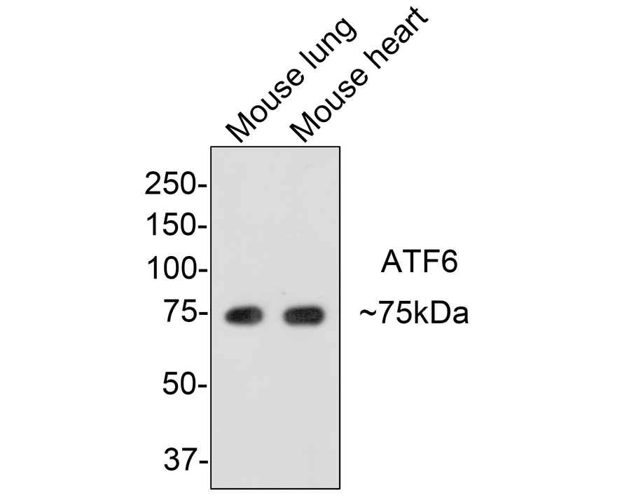
Fig2: Western blot analysis of ATF6 on different lysates with Mouse anti-ATF6 antibody (EM1701-94) at 1/500 dilution.
Lane 1: Mouse lung tissue lysate (20 µg/Lane)
Lane 2: Mouse heart tissue lysate (20 µg/Lane)
Predicted band size: 75 kDa
Observed band size: 75 kDa
Exposure time: 2 minutes;
8% SDS-PAGE gel.
Proteins were transferred to a PVDF membrane and blocked with 5% NFDM/TBST for 1 hour at room temperature. The primary antibody (EM1701-94) at 1/500 dilution was used in 5% NFDM/TBST at room temperature for 2 hours. Goat Anti-Mouse IgG - HRP Secondary Antibody (HA1006) at 1:150,000 dilution was used for 1 hour at room temperature.
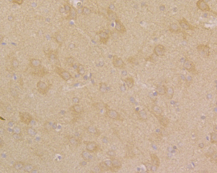
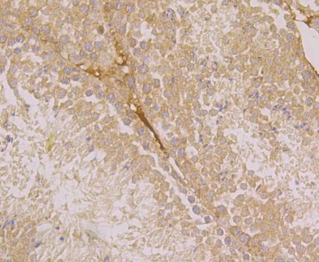
Fig3: Immunohistochemical analysis of paraffin-embedded rat brain tissue using anti-ATF6 antibody. Counter stained with hematoxylin.
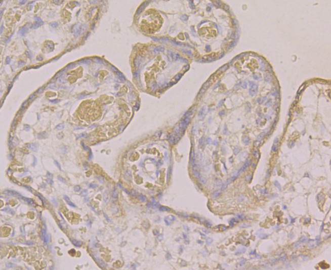
Fig4: Immunohistochemical analysis of paraffin-embedded human placenta tissue using anti-ATF6 antibody. Counter stained with hematoxylin.
Fig5: Immunohistochemical analysis of paraffin-embedded mouse testis tissue using anti-ATF6 antibody. Counter stained with hematoxylin.
Fig6: Flow cytometric analysis of HepG2 cells with ATF6 antibody at 1/50 dilution (pink purple) compared with an unlabelled control (cells without incubation with primary antibody; yellow). Alexa Fluor 488-conjugated goat anti-mouse IgG was used as the secondary antibody.
背景文献
1. Kohl S et al. Mutations in the unfolded protein response regulator ATF6 cause the cone dysfunction disorder achromatopsia. Nat Genet 47:757-765 (2015).
2. Lynch J M et al. A thrombospondin-dependent pathway for a protective ER stress response. Cell 149:1257-1268 (2012).
 会员登录
会员登录 购物车()
购物车()

 成功收藏产品


