产品描述Serine/threonine-protein kinase and endoribonuclease that acts as a key sensor for the endoplasmic reticulum unfolded protein response (UPR). In unstressed cells, the endoplasmic reticulum luminal domain is maintained in its inactive monomeric state by binding to the endoplasmic reticulum chaperone HSPA5/BiP. Accumulation of misfolded protein in the endoplasmic reticulum causes release of HSPA5/BiP, allowing the luminal domain to homodimerize, promoting autophosphorylation of the kinase domain and subsequent activation of the endoribonuclease activity . The endoribonuclease activity is specific for XBP1 mRNA and excises 26 nucleotides from XBP1 mRNA . The resulting spliced transcript of XBP1 encodes a transcriptional activator protein that up-regulates expression of UPR target genes. Acts as an upstream signal for ER stress-induced GORASP2-mediated unconventional (ER/Golgi-independent) trafficking of CFTR to cell membrane by modulating the expression and localization of SEC16A .
产品名称Anti-ERN1 antibody
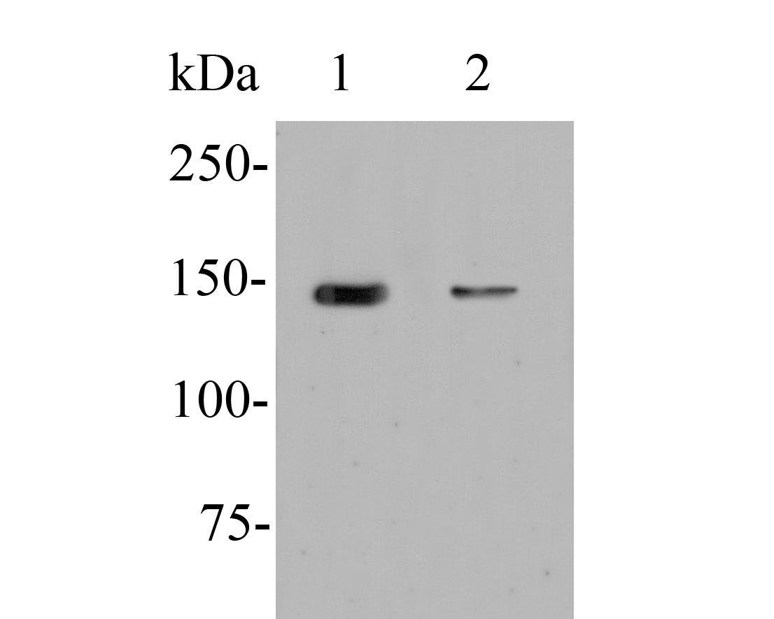
分子量Predicted band size 110 kDa.
种属反应性Human,Rat
验证应用WB,IHC-P
抗体类型兔多抗
免疫原Synthetic peptide within N-terminal Human ERN1.
偶联Non-conjugated
性能
形态Liquid
浓度1 mg/ml.
存放说明Store at +4℃ after thawing. Aliquot store at -20℃. Avoid repeated freeze / thaw cycles.
Fig1: Western blot analysis of ERN1 on different lysates. Proteins were transferred to a PVDF membrane and blocked with 5% BSA in PBS for 1 hour at room temperature. The primary antibody (ER1902-90, 1/500) was used in 5% BSA at room temperature for 2 hours. Goat Anti-Rabbit IgG - HRP Secondary Antibody (HA1001) at 1:5,000 dilution was used for 1 hour at room temperature.
Positive control:
Lane 1: C2C12 cell lysate
Lane 2: HepG2 cell lysate
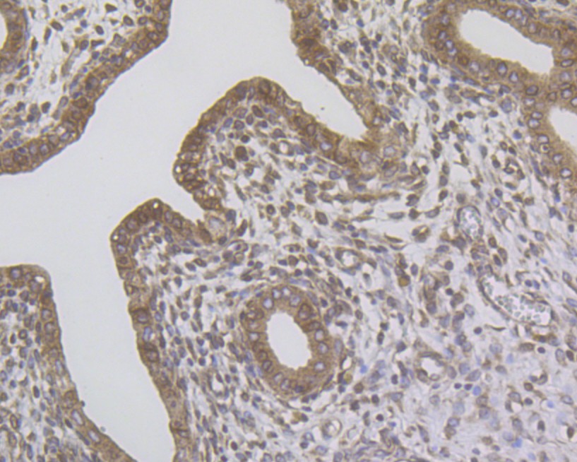
Fig2: Immunohistochemical analysis of paraffin-embedded rat uterus tissue using anti-ERN1 antibody. The section was pre-treated using heat mediated antigen retrieval with Tris-EDTA buffer (pH 8.0-8.4) for 20 minutes.The tissues were blocked in 5% BSA for 30 minutes at room temperature, washed with ddH2O and PBS, and then probed with the primary antibody (ER1902-90, 1/100) for 30 minutes at room temperature. The detection was performed using an HRP conjugated compact polymer system. DAB was used as the chromogen. Tissues were counterstained with hematoxylin and mounted with DPX.
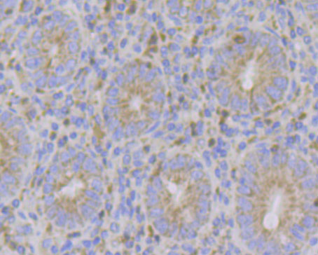
Fig3: Immunohistochemical analysis of paraffin-embedded human appendix tissue using anti-ERN1 antibody. The section was pre-treated using heat mediated antigen retrieval with Tris-EDTA buffer (pH 8.0-8.4) for 20 minutes.The tissues were blocked in 5% BSA for 30 minutes at room temperature, washed with ddH2O and PBS, and then probed with the primary antibody (ER1902-90, 1/100) for 30 minutes at room temperature. The detection was performed using an HRP conjugated compact polymer system. DAB was used as the chromogen. Tissues were counterstained with hematoxylin and mounted with DPX.
背景文献
1. Eletto D.et.al.Protein disulfide isomerase A6 controls the decay of IRE1alpha signaling via disulfide-dependent association.Mol. Cell 53:562-576(2014).
 会员登录
会员登录 购物车()
购物车()

 成功收藏产品


