 会员登录
会员登录 购物车()
购物车()

- 商城价格 登录后可查看价格
- 货号 ET1608-56
- 品牌 huabio/华安生物 ( 经销商 )
- CAS号
- 规格/包装 50ul
- 单位 支
- 储存条件 -20℃
- 现货状态 一周
-
数量
- +
 成功收藏产品
 - 立即购买
 热门推荐
热门推荐
-
概述
- 产品描述The main role of PARP (found in the cell nucleus) is to detect and initiate an immediate cellular response to metabolic, chemical, or radiation-induced single-strand DNA breaks (SSB) by signaling the enzymatic machinery involved in the SSB repair. Once PARP detects a SSB, it binds to the DNA, undergoes a structural change, and begins the synthesis of a polymeric adenosine diphosphate ribose (poly (ADP-ribose) or PAR) chain, which acts as a signal for the other DNA-repairing enzymes. Target enzymes include DNA ligase III (LigIII), DNA polymerase beta (polβ), and scaffolding proteins such as X-ray cross-complementing gene 1 (XRCC1). After repairing, the PAR chains are degraded via Poly(ADP-ribose) glycohydrolase (PARG). PARP enzymes are essential in a number of cellular functions, including expression of inflammatory genes: PARP1 is required for the induction of ICAM-1 gene expression by cardiac myocytes and smooth muscle cells, in response to TNF.
- 产品名称Anti-PARP Recombinant Rabbit Monoclonal Antibody [SU03-68]
- 分子量Predicted band size: 113 kDa
- 种属反应性Human,Mouse, Rat
- 验证应用WB,ICC/IF,IHC-P,FC
- 抗体类型重组兔单抗
- 免疫原Synthetic peptide within N-terminal human PARP.
- 偶联Non-conjugated
-
性能
- 形态Liquid
- 浓度1 mg/mL.
- 存放说明Store at +4℃ after thawing. Aliquot store at -20℃ or -80℃. Avoid repeated freeze / thaw cycles.
- 存储缓冲液1*TBS (pH7.4), 0.05% BSA, 40% Glycerol. Preservative: 0.05% Sodium Azide.
- 亚型IgG
- 纯化方式Protein A affinity purified.
- 亚细胞定位Nucleus, nucleolus, chromosome.
-
数据链接SwissProt: P09874 Human
SwissProt: P11103 Mouse
SwissProt: P27008 Rat
-
其它名称
- ADP ribosyltransferase (NAD+; poly (ADP ribose) polymerase) antibody
- ADP ribosyltransferase diphtheria toxin like 1 antibody
- ADP ribosyltransferase NAD(+) antibody
-
应用
WB: 1:1,000
ICC/IF: 1:50-1:200
IHC-P: 1:500
FC: 1:50-1:100
-

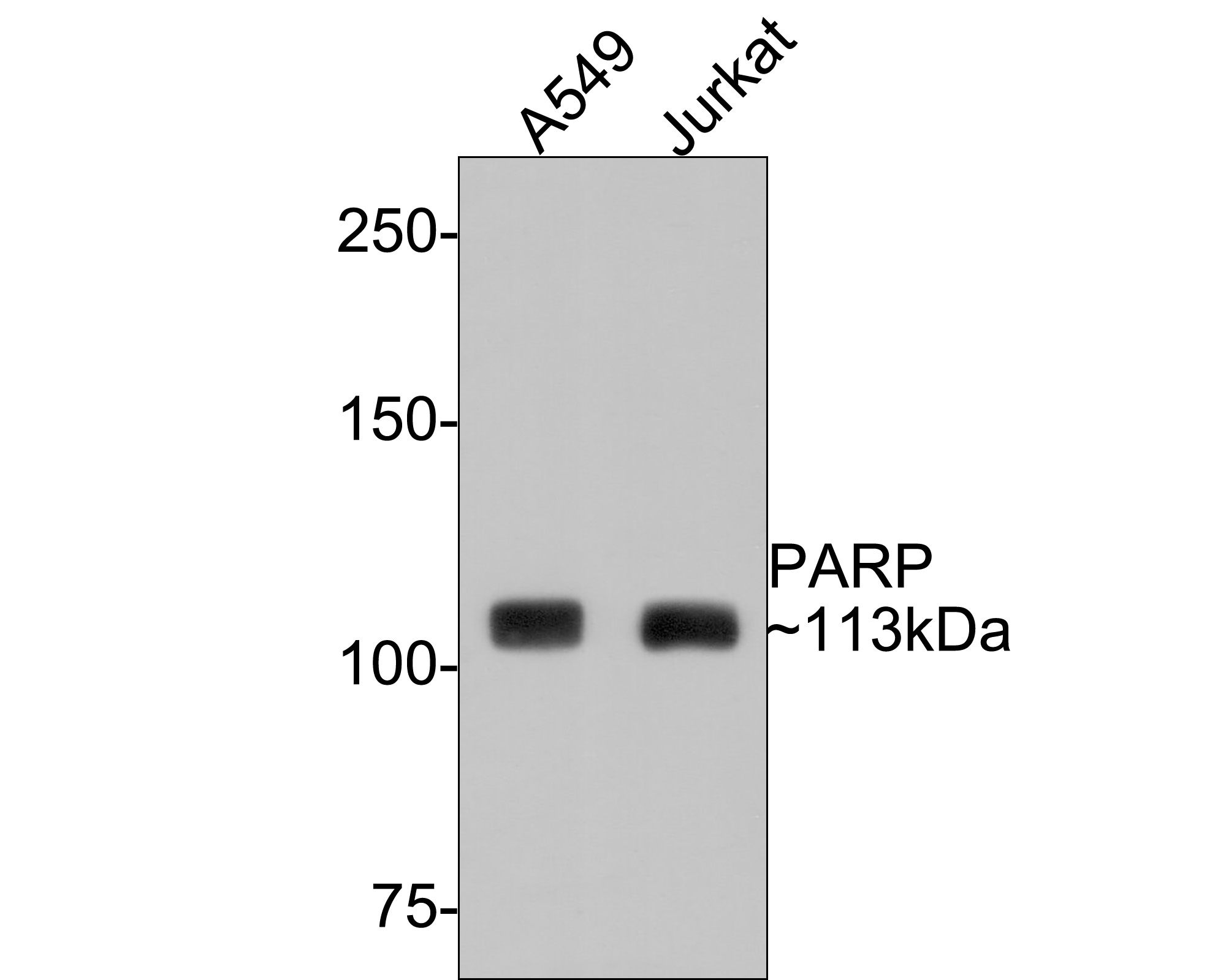
Fig1: Western blot analysis of PARP on different lysates with Rabbit anti-PARP antibody (ET1608-56) at 1/500 dilution.
Lane 1: A549 cell lysate
Lane 2: Jurkat cell lysate
Lysates/proteins at 10 µg/Lane.
Predicted band size: 113 kDa
Observed band size: 113 kDa
Exposure time: 1 minute;
6% SDS-PAGE gel.
Proteins were transferred to a PVDF membrane and blocked with 5% NFDM/TBST for 1 hour at room temperature. The primary antibody (ET1608-56) at 1/500 dilution was used in 5% NFDM/TBST at room temperature for 2 hours. Goat Anti-Rabbit IgG - HRP Secondary Antibody (HA1001) at 1:300,000 dilution was used for 1 hour at room temperature.
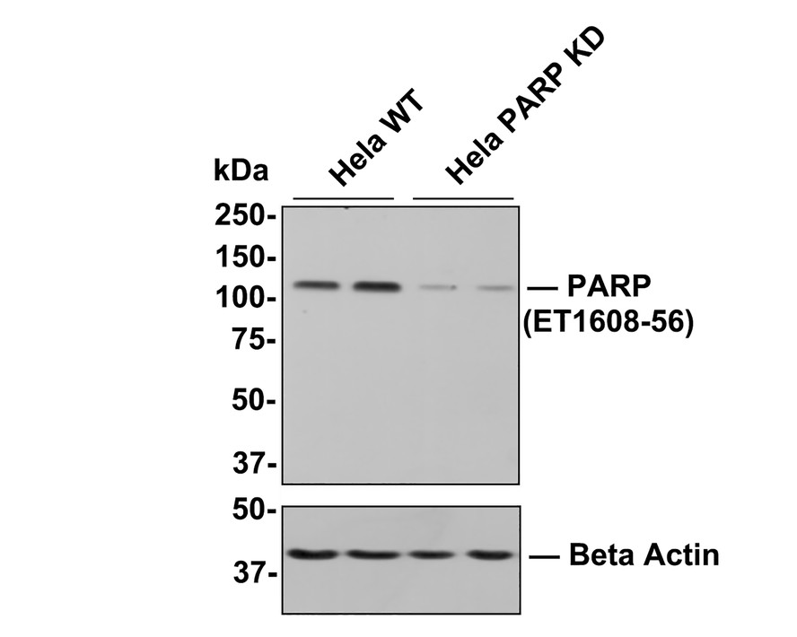
Fig2: All lanes: Western blot analysis of PARP with anti-PARP antibody[SU03-68] (ET1608-56) at 1:500 dilution.
Lane 1/2: Wild-type Hela whole cell lysate (10 µg).
Lane 3/4: PARP knockdown Hela whole cell lysate (10 µg).
ET1608-56 was shown to specifically react with PARP in wild-type Hela cells. Weakened bands were observed when PARP knockdown samples were tested. Wild-type and PARP knockdown samples were subjected to SDS-PAGE. Proteins were transferred to a PVDF membrane and blocked with 5% NFDM in TBST for 1 hour at room temperature. The primary antibody (ET1608-56, 1/500) was used in 5% BSA at room temperature for 2 hours. Goat Anti-Rabbit IgG-HRP Secondary Antibody (HA1001) at 1:200,000 dilution was used for 1 hour at room temperature.
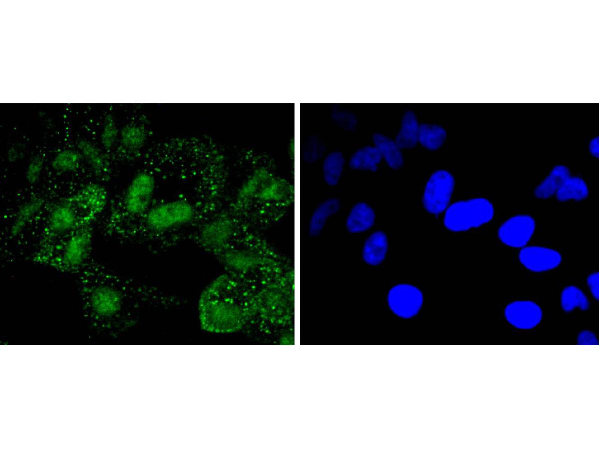
Fig3: ICC staining of PARP in Hela cells (green). Formalin fixed cells were permeabilized with 0.1% Triton X-100 in TBS for 10 minutes at room temperature and blocked with 1% Blocker BSA for 15 minutes at room temperature. Cells were probed with the primary antibody (ET1608-56, 1/50) for 1 hour at room temperature, washed with PBS. Alexa Fluor®488 Goat anti-Rabbit IgG was used as the secondary antibody at 1/1,000 dilution. The nuclear counter stain is DAPI (blue).

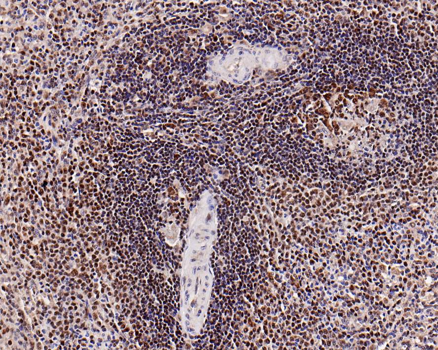
Fig4: Immunohistochemical analysis of paraffin-embedded human spleen tissue with Rabbit anti-PARP antibody (ET1608-56) at 1/500 dilution.
The section was pre-treated using heat mediated antigen retrieval with Tris-EDTA buffer (pH 9.0) for 20 minutes. The tissues were blocked in 1% BSA for 20 minutes at room temperature, washed with ddH2O and PBS, and then probed with the primary antibody (ET1608-56) at 1/500 dilution for 1 hour at room temperature. The detection was performed using an HRP conjugated compact polymer system. DAB was used as the chromogen. Tissues were counterstained with hematoxylin and mounted with DPX.
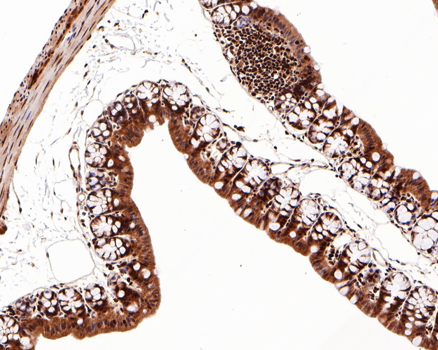
Fig5: Immunohistochemical analysis of paraffin-embedded mouse large intestine tissue with Rabbit anti-PARP antibody (ET1608-56) at 1/500 dilution.
The section was pre-treated using heat mediated antigen retrieval with Tris-EDTA buffer (pH 9.0) for 20 minutes. The tissues were blocked in 1% BSA for 20 minutes at room temperature, washed with ddH2O and PBS, and then probed with the primary antibody (ET1608-56) at 1/500 dilution for 1 hour at room temperature. The detection was performed using an HRP conjugated compact polymer system. DAB was used as the chromogen. Tissues were counterstained with hematoxylin and mounted with DPX.
Fig6: Flow cytometric analysis of PARP was done on Hela cells. The cells were fixed, permeabilized and stained with the primary antibody (ET1608-56, 1/50) (red). After incubation of the primary antibody at room temperature for an hour, the cells were stained with a Alexa Fluor 488-conjugated Goat anti-Rabbit IgG Secondary antibody at 1/1000 dilution for 30 minutes.Unlabelled sample was used as a control (cells without incubation with primary antibody; black).

Fig7: Immunohistochemical analysis of paraffin-embedded human breast carcinoma tissue with Rabbit anti-PARP antibody (ET1608-56) at 1/500 dilution.
The section was pre-treated using heat mediated antigen retrieval with Tris-EDTA buffer (pH 9.0) for 20 minutes. The tissues were blocked in 1% BSA for 20 minutes at room temperature, washed with ddH2O and PBS, and then probed with the primary antibody (ET1608-56) at 1/500 dilution for 1 hour at room temperature. The detection was performed using an HRP conjugated compact polymer system. DAB was used as the chromogen. Tissues were counterstained with hematoxylin and mounted with DPX.


-
背景文献
-
1. Cao C et al. The long intergenic noncoding RNA UFC1, a target of MicroRNA 34a, interacts with the mRNA stabilizing protein HuR to increase levels of -catenin in HCC cells. Gastroenterology 148:415-26.e18 (2015).
2. Gao S et al. Ischemia-reperfusion injury of the retina is linked to necroptosis via the ERK1/2-RIP3 pathway. Mol Vis 20:1374-87 (2014).