产品描述The expression of DUSP1 gene is induced in human skin fibroblasts by oxidative/heat stress and growth factors. It specifies a protein with structural features similar to members of the non-receptor-type protein-tyrosine phosphatase family, and which has significant amino-acid sequence similarity to a Tyr/Ser-protein phosphatase encoded by the late gene H1 of vaccinia virus. The bacterially expressed and purified DUSP1 protein has intrinsic phosphatase activity, and specifically inactivates mitogen-activated protein (MAP) kinase in vitro by the concomitant dephosphorylation of both its phosphothreonine and phosphotyrosine residues. Furthermore, it suppresses the activation of MAP kinase by oncogenic ras in extracts of Xenopus oocytes. Thus, DUSP1 may play an important role in the human cellular response to environmental stress as well as in the negative regulation of cellular proliferation.
WB: 1:500-1:2,000
IHC-P: 1:50-1:200
FC: 1:50-1:100
IP: Use at an assay dependent concentration.
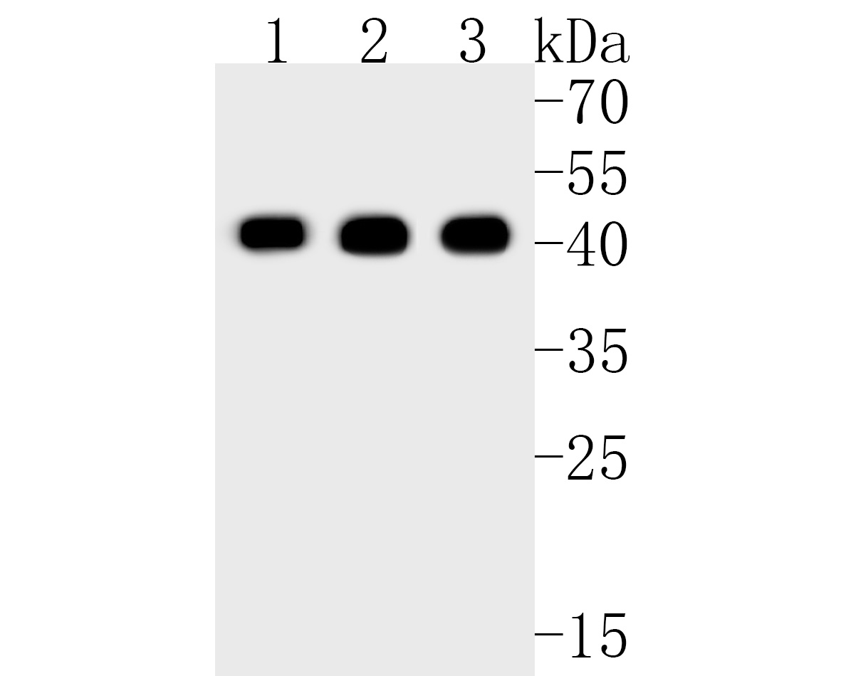
Fig1: Western blot analysis of DUSP1 on different lysates. Proteins were transferred to a PVDF membrane and blocked with 5% BSA in PBS for 1 hour at room temperature. The primary antibody (ET1701-82, 1/500) was used in 5% BSA at room temperature for 2 hours. Goat Anti-Rabbit IgG - HRP Secondary Antibody (HA1001) at 1:5,000 dilution was used for 1 hour at room temperature.
Positive control:
Lane 1: NIH/3T3 cell lysate
Lane 2: Jurkat cell lysate
Lane 3: mouse spleen tissue lysate
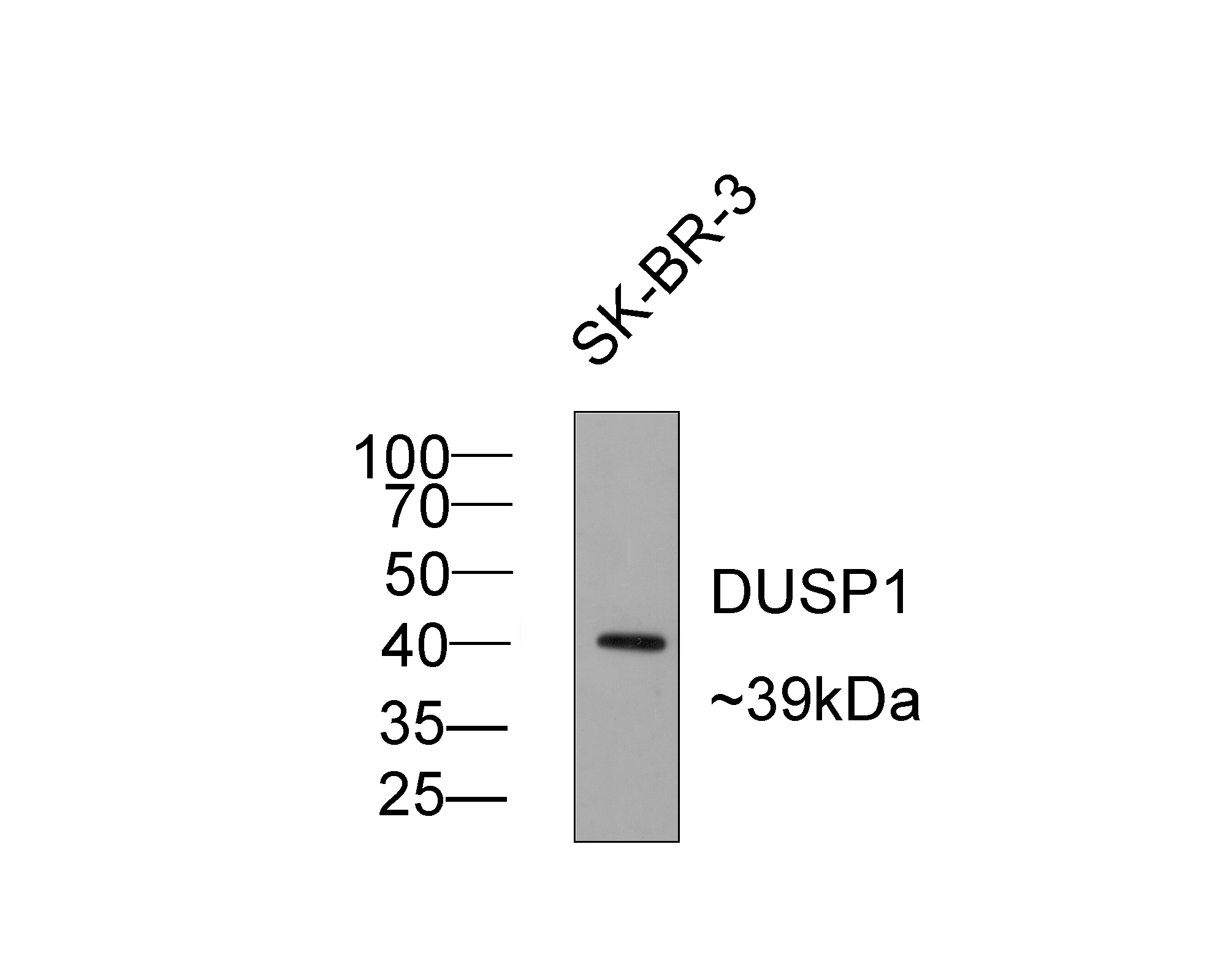
Fig2: Western blot analysis of DUSP1 on SK-BR-3 cell tissue lysates with Rabbit anti-DUSP1 antibody (ET1701-82) at 1/500 dilution.
Lysates/proteins at 10 µg/Lane.
Predicted band size:39 kDa
Observed band size: 39 kDa
Exposure time: 2 minutes;
10% SDS-PAGE gel.
Proteins were transferred to a PVDF membrane and blocked with 5% NFDM/TBST for 1 hour at room temperature. The primary antibody (ET1701-82) at 1/500 dilution was used in 5% NFDM/TBST at room temperature for 2 hours. Goat Anti-Rabbit IgG - HRP Secondary Antibody (HA1001) at 1:5,000 dilution was used for 1 hour at room temperature.
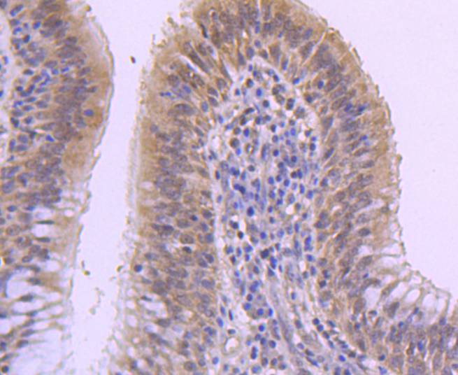
Fig3: Immunohistochemical analysis of paraffin-embedded human lung carcinoma tissue using anti-DUSP1 antibody. The section was pre-treated using heat mediated antigen retrieval with Tris-EDTA buffer (pH 8.0-8.4) for 20 minutes.The tissues were blocked in 5% BSA for 30 minutes at room temperature, washed with ddH2O and PBS, and then probed with the primary antibody (ET1701-82, 1/50) for 30 minutes at room temperature. The detection was performed using an HRP conjugated compact polymer system. DAB was used as the chromogen. Tissues were counterstained with hematoxylin and mounted with DPX.
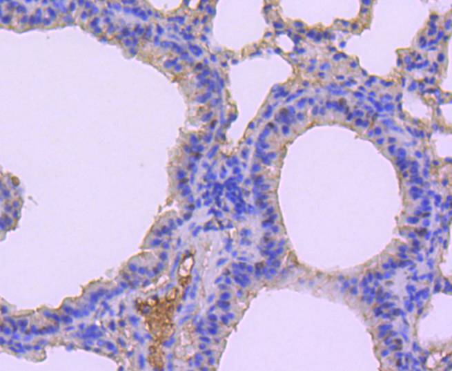
Fig4: Immunohistochemical analysis of paraffin-embedded mouse lung tissue using anti-DUSP1 antibody. The section was pre-treated using heat mediated antigen retrieval with Tris-EDTA buffer (pH 8.0-8.4) for 20 minutes.The tissues were blocked in 5% BSA for 30 minutes at room temperature, washed with ddH2O and PBS, and then probed with the primary antibody (ET1701-82, 1/50) for 30 minutes at room temperature. The detection was performed using an HRP conjugated compact polymer system. DAB was used as the chromogen. Tissues were counterstained with hematoxylin and mounted with DPX.
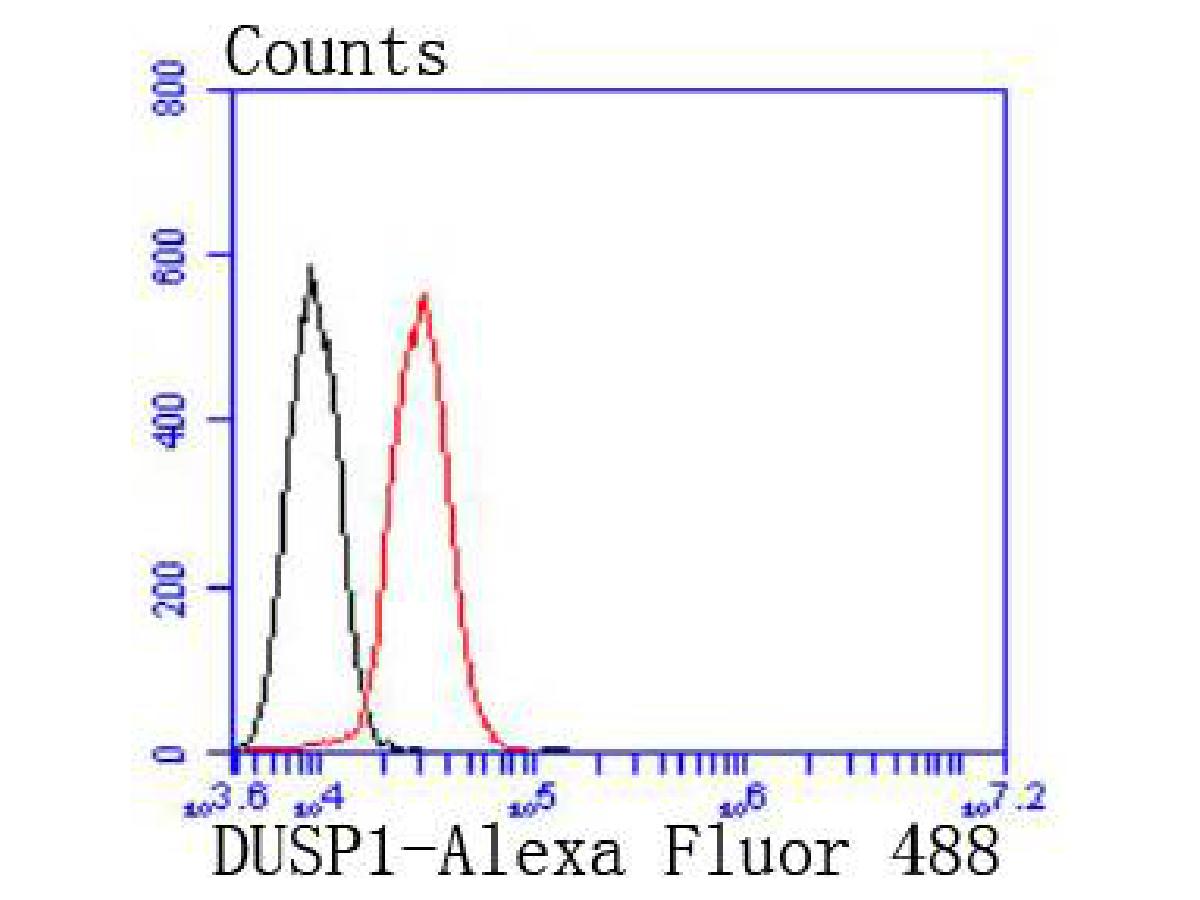
Fig5: Flow cytometric analysis of DUSP1 was done on HepG2 cells. The cells were fixed, permeabilized and stained with the primary antibody (ET1701-82, 1/50) (red). After incubation of the primary antibody at room temperature for an hour, the cells were stained with a Alexa Fluor 488-conjugated Goat anti-Rabbit IgG Secondary antibody at 1/1000 dilution for 30 minutes.Unlabelled sample was used as a control (cells without incubation with primary antibody; black).
背景文献
1. Szelenyi ER & Urso ML Time-course analysis of injured skeletal muscle suggests a critical involvement of ERK1/2 signaling in the acute inflammatory response. Muscle Nerve 45:552-61 (2012).
2. Asrih M et al. Role of ERK1/2 activation in microtubule stabilization and glucose transport in cardiomyocytes. Am J Physiol Endocrinol Metab 301:E836-43 (2011).
 会员登录
会员登录 购物车()
购物车()

 成功收藏产品


