产品描述Endothelial PAS domain-containing protein 1 (EPAS1, also known as hypoxia-inducible factor-2alpha (HIF-2α)) is a protein that is encoded by the EPAS1 gene in mammals. It is a type of hypoxia-inducible factor, a group of transcription factors involved in the physiological response to oxygen concentration. The gene is active under hypoxic conditions. It is also important in the development of the heart, and for maintaining the catecholamine balance required for protection of the heart. Mutation often leads to neuroendocrine tumors. The EPAS1 gene encodes one subunit of a transcription factor involved in the induction of genes regulated by oxygen, and which is induced as oxygen concentration falls (hypoxia). The protein contains a basic helix-loop-helix protein dimerization domain as well as a domain found in signal transduction proteins which respond to oxygen levels. EPAS1 is involved in the development of the embryonic heart and is expressed in endothelial cells that line the walls of blood vessels in the umbilical cord. EPAS1 is also essential for the maintenance of catecholamine homeostasis and protection against heart failure during early embryonic development. Catecholamines regulated by EPAS1 include epinephrine and norepinephrine. It is critical that the production of catecholamines remain in homeostatic conditions so that both the delicate fetal heart and the adult heart do not overexert themselves and induce heart failure. Catecholamine production in the embryo is related to control of cardiac output by increasing the fetal heart rate.
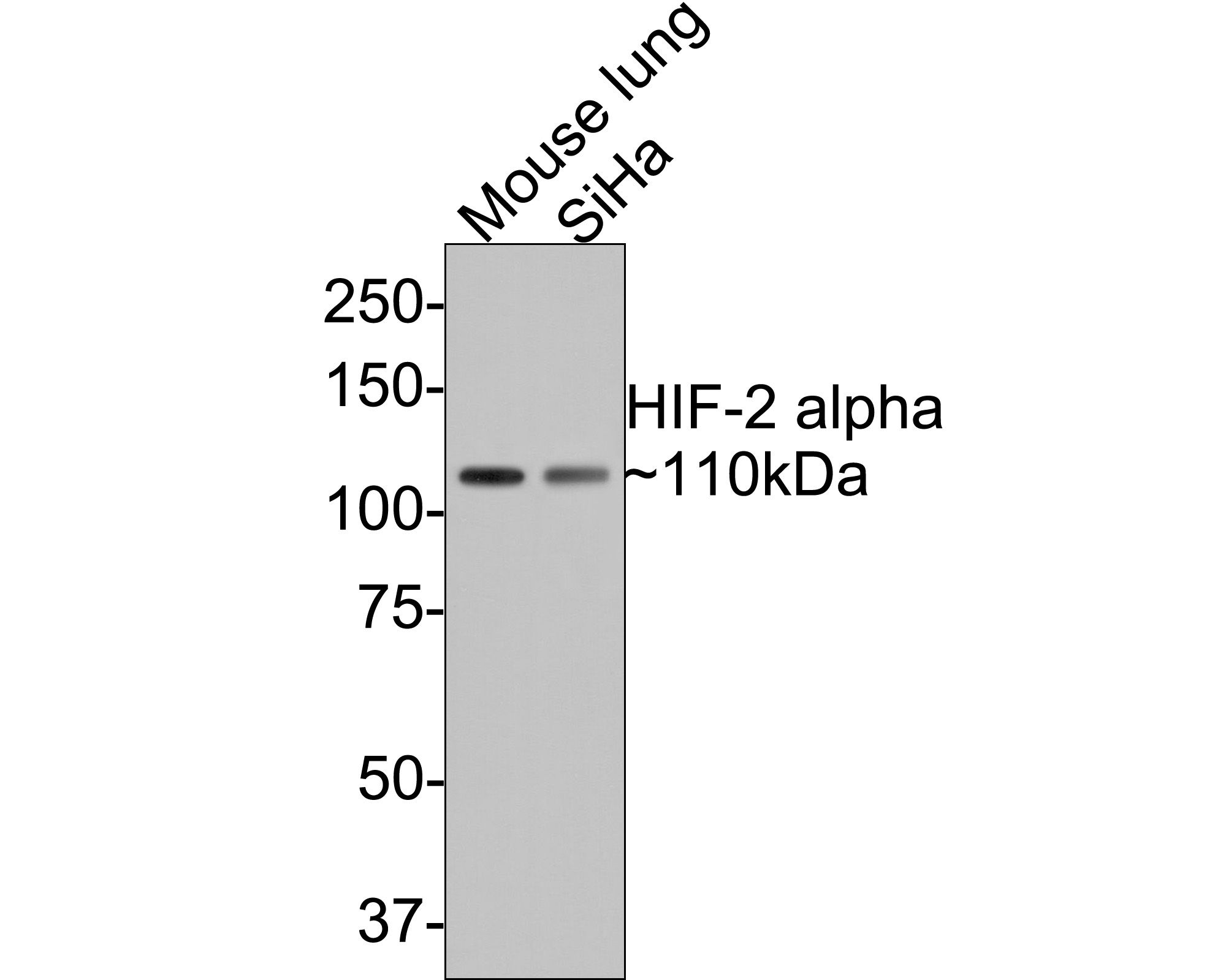
Fig1: Western blot analysis of HIF-2 alpha on different lysates with Rabbit anti-HIF-2 alpha antibody (ET7107-32) at 1/500 dilution.
Lane 1: Mouse lung tissue lysate (20 µg/Lane)
Lane 2: Siha cell lysate (10 µg/Lane)
Predicted band size: 96 kDa
Observed band size: 110 kDa
Exposure time: 1 minute;
8% SDS-PAGE gel.
Proteins were transferred to a PVDF membrane and blocked with 5% NFDM/TBST for 1 hour at room temperature. The primary antibody (ET7107-32) at 1/500 dilution was used in 5% NFDM/TBST at room temperature for 2 hours. Goat Anti-Rabbit IgG - HRP Secondary Antibody (HA1001) at 1:300,000 dilution was used for 1 hour at room temperature.
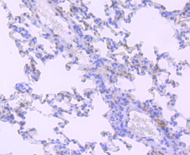
Fig2: Immunohistochemical analysis of paraffin-embedded rat lung tissue using anti-HIF-2 alpha antibody. Counter stained with hematoxylin.
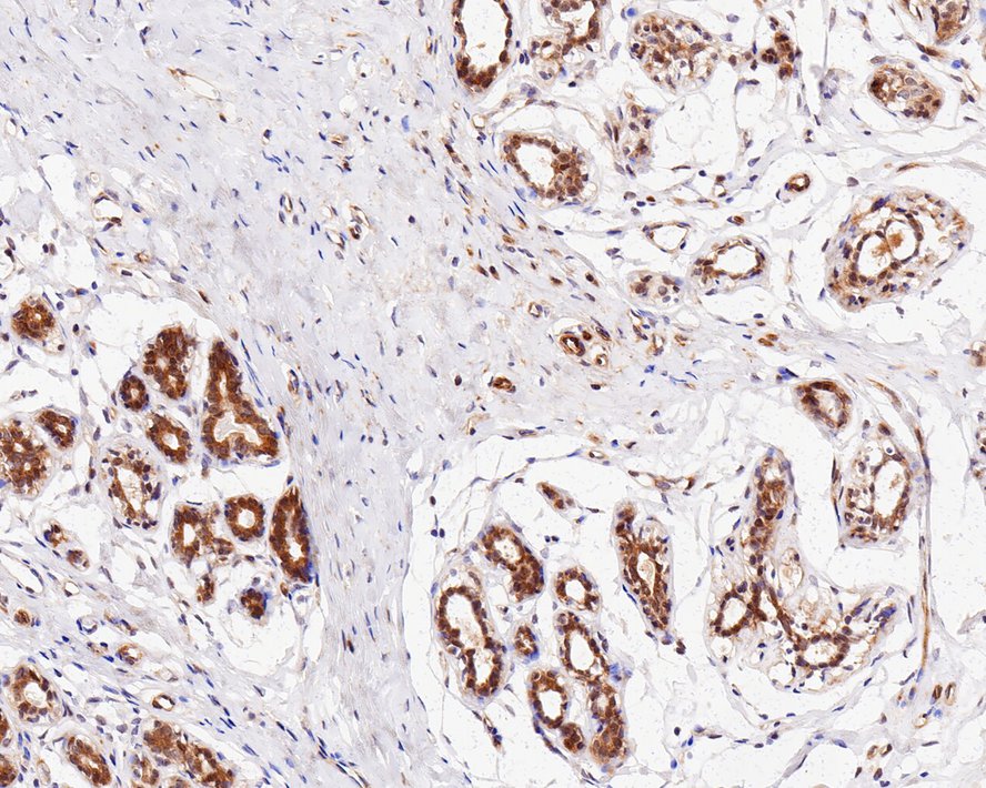
Fig3: Immunohistochemical analysis of paraffin-embedded human breast tissue with Rabbit anti-HIF-2 alpha antibody (ET7107-32) at 1/1,000 dilution.
The section was pre-treated using heat mediated antigen retrieval with sodium citrate buffer (pH 6.0) for 2 minutes. The tissues were blocked in 1% BSA for 20 minutes at room temperature, washed with ddH2O and PBS, and then probed with the primary antibody (ET7107-32) at 1/1,000 dilution for 1 hour at room temperature. The detection was performed using an HRP conjugated compact polymer system. DAB was used as the chromogen. Tissues were counterstained with hematoxylin and mounted with DPX.
Fig4: Immunohistochemical analysis of paraffin-embedded human placenta tissue using anti-HIF-2 alpha antibody. Counter stained with hematoxylin.
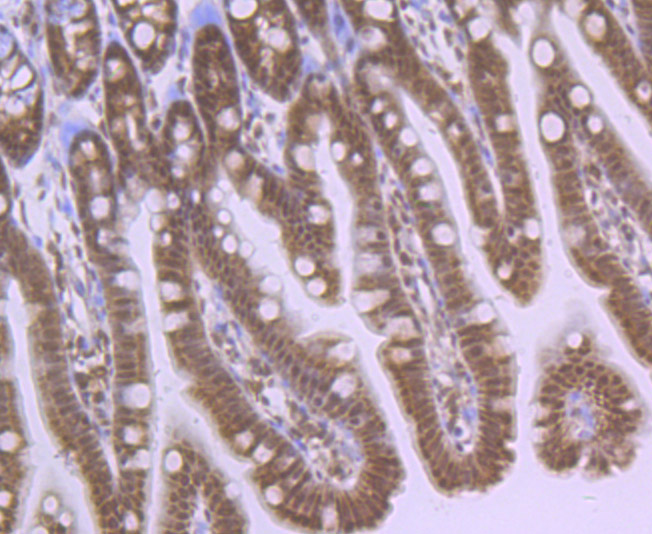
Fig5: Immunohistochemical analysis of paraffin-embedded mouse colon tissue using anti-HIF-2 alpha antibody. Counter stained with hematoxylin.
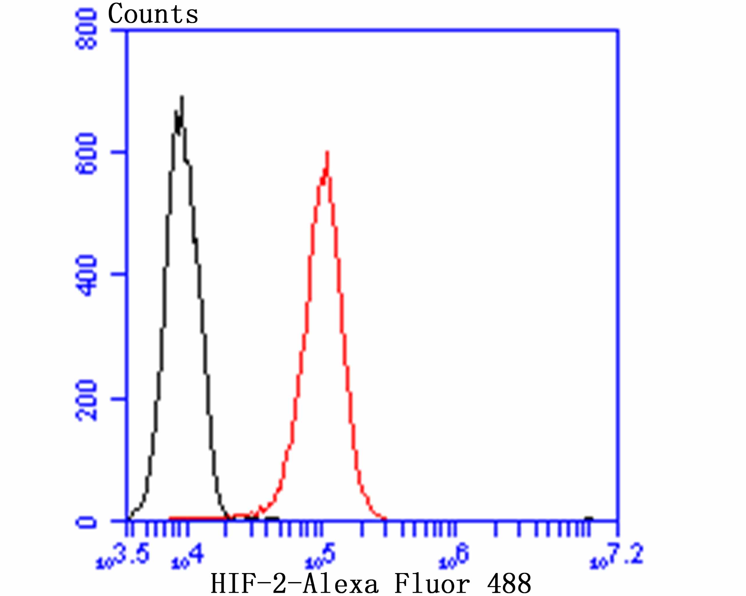
Fig6: Flow cytometric analysis of HUVEC cells with HIF-2 antibody at 1/100 dilution (red) compared with an unlabelled control (cells without incubation with primary antibody; black). Alexa Fluor 488-conjugated goat anti rabbit IgG was used as the secondary antibody.
背景文献
1. Ema M et al. Molecular mechanisms of transcription activation by HLF and HIF1alpha in response to hypoxia: their stabilization and redox signal-induced interaction with CBP/p300. EMBO J 18:1905-1914 (1999).
2. Furlow P W et al. Erythrocytosis-associated HIF-2alpha mutations demonstrate a critical role for residues C-terminal to the hydroxylacceptor proline. J Biol Chem 284:9050-9058 (2009).
 会员登录
会员登录 购物车()
购物车()

 成功收藏产品


